AnatomI 3D is a application based on Virtual Reality (VR) which offers interactive visualization of 3D human body structures associated to explanatory texts. It allows the visualization of any anatomical structure provided by the user and offers the individual or group selection of these structures (Figure 1).
Through te import of several 3D models and their respective description it is possible to use the AnatomI 3D to study any part of t human body. The models and texts must be provided by the user. The AnatomI 3D has a set of menus for interactive dissection and control of transparency levels for each model of the structure displayed (Figure 2A and 2B).
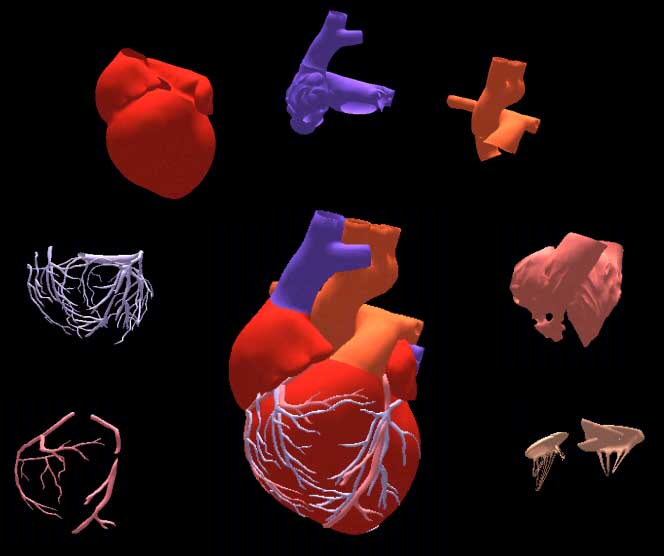
Figure 1: Example of 3D models used by the AnatomI 3D to compose the human heart.
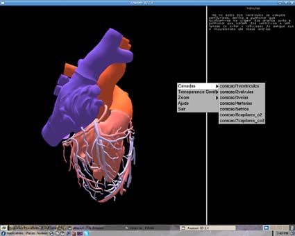
Figure 2: System in operation: menus to control the models displayed (dissection) and the transparency levels of each one. The right side of the screen presents the text related to a model selected by the user.
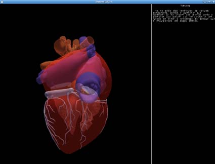
Figure 2: System in operation: menus to control the models displayed (dissection) and the transparency levels of each one. The right side of the screen presents the text related to a model selected by the user.
The system was developed to run in the VirtWall platform, a visualization system based on free tools and low-cost hardware. This platform allows multiple user visualization, what is useful for classes. This feature offers stereoscopy visualization and favor discussions related to the structure observed. Besides this platform, the system also run on ordinary PCs. In this case the visualization can monoscopic or the anagliph mode can be selected for stereoscopic visualization. (Figure 3).
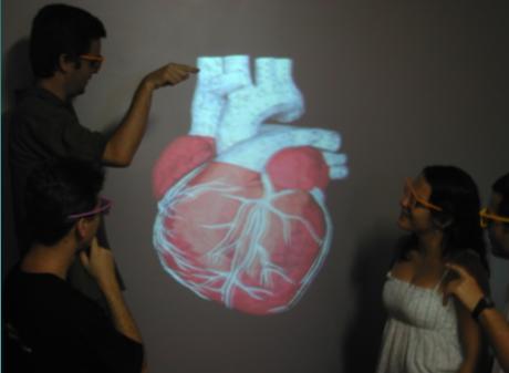
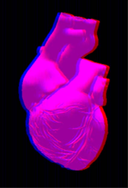
Figure 3: The AnatomI 3D running in the VirtWall platform and in the anagliph mode for PCs.
Technical Information
The AnatomI 3D development was completely based on free tools. Its implementation used the C++ programming language, the OpenGL graphic library and the Fedora Linux 3.0 distribution.
The data structure (DS) of the AnatomI 3D is based in a Corner-Table (Cunha et al. 2006) and support the use of triangular meshes.
3D models designed in any modelling package can be used by the system. In this case, they must be saved in the VRML 1.0 or 2.0 format (.WRL) to be recognized by the AnatomI 3D (Figure 4).
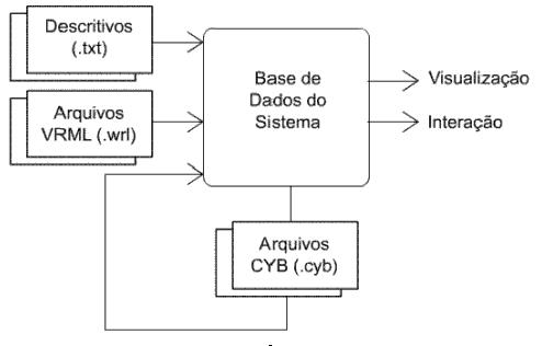
Figure 4: Basic diagram of the AnatomI 3D.
To allow a fast load of the models, the AnatomI 3D has a pre-processed format with .CYB extension. The pre-processed files are created at the first load of the models. Later, the user can use these new files, composed by the models and other parameters used by the system, to accelerate the charge of the AnatomI 3D.
The .CYB files also contain information used to lighting. It is calculated at the first load of the models. This way, vertex and polygons normals can be used by the lighting method to remove discontinuity between the polygons that compose each model.
 Return